

Most ACL injury biomechanics research comes from video analysis, cadaver studies, or biomechanical modelling.
But this study captured two ACL ruptures occurring during 3D motion capture and force plate testing.
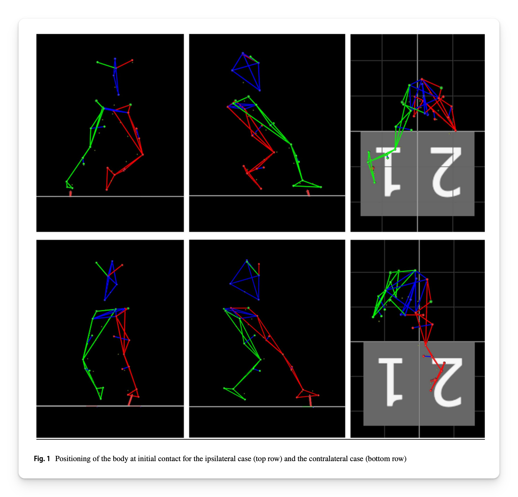
Both athletes were adolescent females undergoing testing following ACL reconstruction. One re-ruptured her reconstructed ACL at 24 weeks post-op. The other ruptured the ACL of her opposite leg at 38 weeks post-op.
Both injuries occurred during an unplanned 90° cutting trial.
Even more interesting, both athletes successfully completed an unplanned cut immediately beforehand which allowed researchers to compare a successful cut with an ACL injury cut within the same athlete.
When two athletes tear their ACLs, do they look the same?
What Did the Researchers Do?
Athletes approached a force plate over 5 meters before performing a 90° cut. During unplanned trials, a light and buzzer indicated the direction they needed to cut.
Researchers measured:
- Approach velocity and deceleration
- Penultimate-step mechanics
- Step length and touchdown distance
- Trunk, pelvis, hip, knee, and foot positions
- Ground reaction forces
- Knee forces and moments
- Timing of peak forces

The Performance Academy: Gain access to the best performance and rehab minds in the world through premium masterclasses, live and recorded events, plus a network of coaches and therapist around the world. Education, community, and networking under one roof, all at your fingertips.
What Were the Results?
Athlete 1: More Flexed and Wider
During the injury-producing cut, Athlete 1:
- Arrived at initial contact faster, 2.47 vs. 2.29 m/s
- Decelerated less from the penultimate step to initial contact, -4.01 vs. -6.11 m/s²
- Planted farther laterally from her pelvis, 0.364 vs. 0.290 m
- Actually had MORE knee flexion, 41° vs. 36°
- Increased resultant GRF from approximately 3.4× to 4.8× bodyweight
- Approximately doubled her knee abductor moment
- More than tripled her external rotator moment
Her injury was therefore not characterized by a more extended knee. Instead, she arrived faster, reached farther laterally, and experienced greater frontal and transverse-plane knee loading.
Athlete 2: More Extended and Farther Forward
Athlete 2 looked completely different.
During the injury-producing cut, she:
- Reached the penultimate step at 3.11 vs. 1.40 m/s
- Reached initial contact at 2.40 vs. 1.42 m/s
- Increased final step length from 0.499 to 0.768 m
- Planted much farther anterior to her pelvis
- Reduced knee flexion from 60° to just 22°
- Increased resultant GRF from approximately 2.4× to 5.4× bodyweight
- Approximately doubled knee compression force
- More than tripled her knee abductor moment
Her knee remained around 22° of flexion for the first 50 to 60 milliseconds before the subsequent valgus collapse.
What Does This Mean?
The key is that there was no single "wrong" cutting position shared by both ACL injuries.
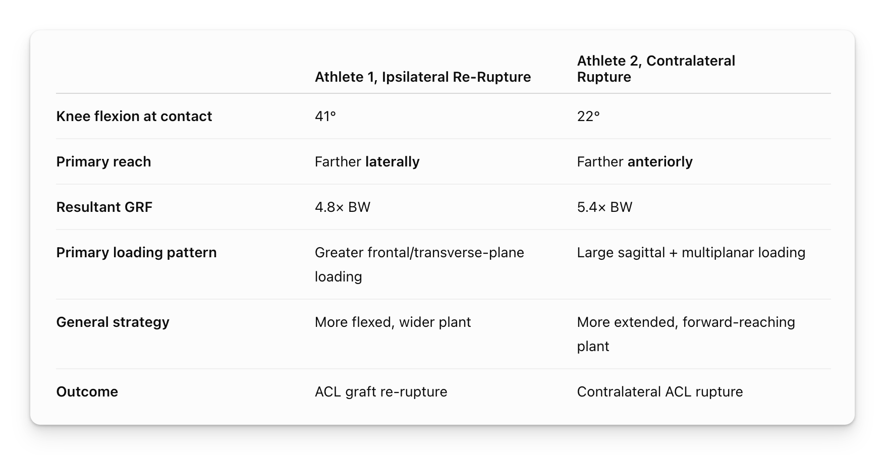
Athlete 1 ⮕ Contacted with 41° of knee flexion, reached farther laterally, and experienced greater frontal and transverse-plane loading.
Athlete 2 ⮕ Contacted with 22° of knee flexion, reached farther anteriorly, and experienced large sagittal and multiplanar loading.
Different positions and strategies leading to ACL failure.
But there were some important similarities upstream of the knee.
During the injury cuts, both athletes:
- Carried more velocity toward the plant
- Did not sufficiently dissipate that additional momentum beforehand
- Used a more outstretched cutting limb
- Experienced substantially greater knee loading
- Had multiple peak forces and moments occur closer together in time
In short, both athletes created a harder momentum problem for the cutting leg to solve. From there, each athlete used a different movement strategy to brake and redirect that momentum.
Limitations
- This was a two-person case series, so these mechanics cannot be considered predictive risk factors.
- Athlete 1 was only 24 weeks post-op and had not yet progressed to running.
- These injuries occurred during laboratory testing rather than competition which gives us compelling data but limits extrapolation to real world environments.
Coach's Takeaway
- There may be no single "wrong" cutting mechanic ⮕ The two athletes demonstrated very different knee positions when their ACLs failed.
- Look upstream from the knee ⮕ Approach velocity and braking strategy influence the momentum the cutting limb eventually has to manage.
- Foot placement needs context ⮕ Both athletes became more outstretched, but one reached laterally while the other reached anteriorly.
- Think in movement strategies, not isolated joint positions ⮕ Knee angle alone doesn't tell the story. Velocity, momentum, foot placement, body orientation, and multiplanar loading all interact.
Perhaps the biggest lesson from these two cases is that different movement strategies can create different pathways to excessive ACL loading.
Instead of searching for the one "bad" cutting position, we may be better served asking, What movement problem is the athlete trying to solve, and how are they solving it?
I hope this helps,
Ramsey
Reference: Kung, S. M., Jackson, M., Withers, D., & Welch, N. (2026). The 3D Biomechanics of Secondary Non-contact ACL Injuries in Two Female Adolescents: A Case Series of an Ipsilateral ACL Re-Rupture and a Contralateral ACL Rupture. Sports Medicine.


