

Patellar tendinopathy is common in jumping sports, with previous research reporting prevalence rates as high as 44.6% among elite volleyball players.
Because symptoms often develop on one side, clinicians may look for structural differences between limbs to explain why one tendon became painful.
Static measurements of femoral, tibial, knee, and ankle alignment are sometimes included in these assessments.
This study examined whether elite volleyball players with unilateral patellar tendinopathy had different frontal-plane skeletal alignment in their symptomatic knee compared with their asymptomatic knee.
Is that limb with patellar tendinopathy structurally aligned differently from the pain-free limb?
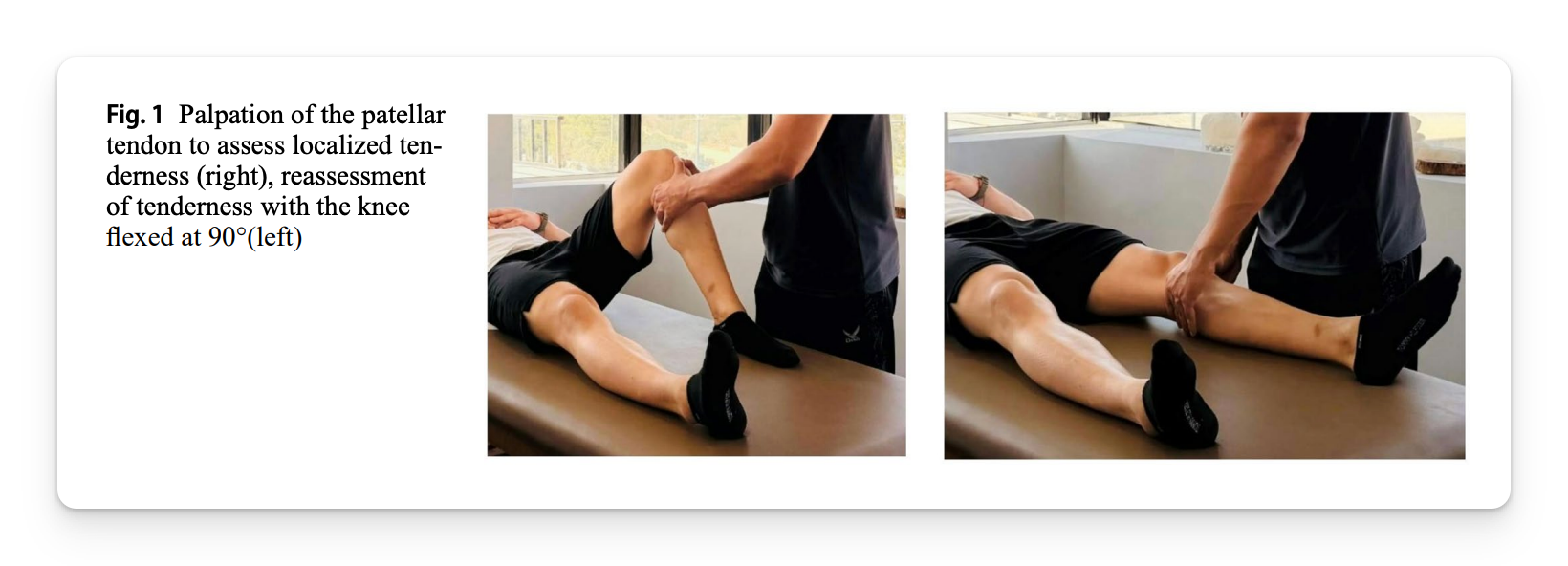
What Did the Researchers Do?
The researchers conducted a cross-sectional study involving 22 elite male volleyball players from Iran:
- Average age: 19.6 years
- Average height: 191.7 cm
- Average body mass: 80.6 kg
- All athletes had unilateral patellar tendinopathy
Each athlete completed standardized, bilateral, weight-bearing radiographs. One blinded examiner measured seven frontal-plane alignment variables across the femur, knee, tibia, and ankle to determine if differences existed between sides.
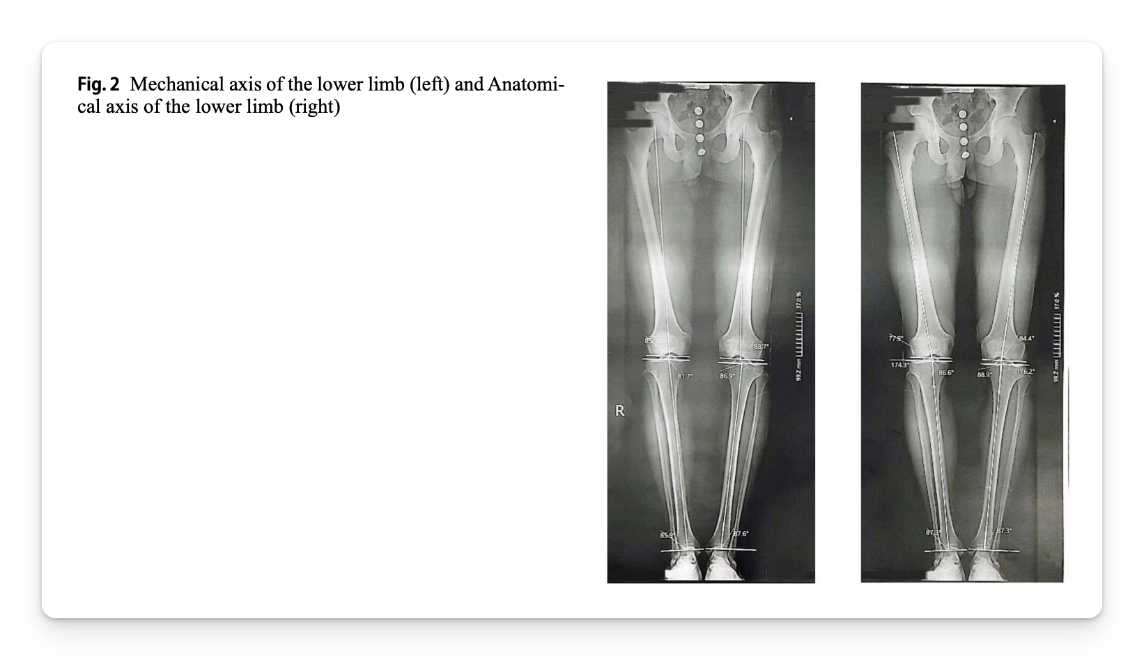
What Were the Results?
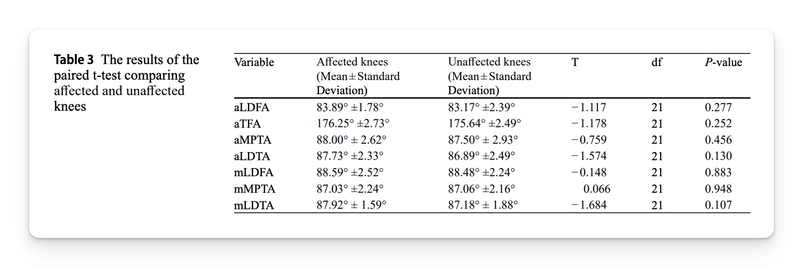
The researchers found no statistically significant differences between the affected and unaffected knees for any of the seven alignment measures.
The actual differences were also small:
- Distal femoral angles differed by less than 1 degree
- Tibiofemoral alignment differed by approximately 0.6 degrees
- Proximal tibial alignment was nearly identical
- Distal tibial alignment differed by less than 1 degree
The painful and pain-free knees were therefore remarkably similar across every static alignment variable measured.
In other words, static lower-limb alignment did not distinguish the knee with patellar tendinopathy from the pain-free knee.
What Does This Mean?
Static frontal-plane alignment did not distinguish the knee with patellar tendinopathy from the pain-free knee and skeletal geometry measured during standing offered. little explanation for which knee had symptoms.
But this is not surprising.
Tendinopathy likely involves interacting factors such as tendon loading, jumping exposure, tendon capacity, recovery, previous symptoms, and individual biological responses.
Limitations
- Only 22 athletes were studied, limiting the ability to detect small differences.
- The cross-sectional design cannot determine whether alignment preceded symptoms.
- Patellar tendinopathy was classified using one clinical test without imaging confirmation.
Coach’s Takeaway
- Do not build an explanation for patellar tendinopathy around a few static alignment measurements.
- Assess symptom behavior, recent workload, jumping exposure, strength, tendon capacity, recovery, and tolerance to progressive loading.
- Use movement testing to understand an athlete’s current function, not to search for one defective pattern that supposedly caused the problem.
I hope this helps,
Ramsey
Reference: Saraee D, Piri H, Sheikhhoseini R, Hajizadeh R. (2026). Comparison of lower limb alignment in knees with and without patellar tendinopathy among elite volleyball players: a cross-sectional study. Journal of Orthopaedic Surgery and Research.


